
The Tumor Board: The Impact of Going Digital
Keith Kaplan, MD, Chief Medical Officer
I have been attending tumor boards on a weekly basis in some capacity or another for over 20 years, since starting my pathology residency. Prior to then, I observed as a medical student on a regular basis. Even if I was on a rotation, like family practice or pediatrics or OB/Gyn, if I had the time, I would try to attend one of the many tumor boards on the hospital campus. I kept the schedule in my PalmPilot Personal Digital Assistant at that time.
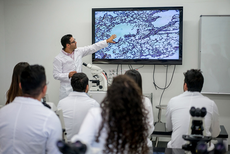
Regardless, since I have been watching, attending or presenting, tumor boards have remained unchanged in terms of their format. Surgeons, medical oncologists, radiation oncologists, radiologists and pathologists gather in a room and discuss a list of cases that came out perhaps a few days before the scheduled meeting. Many subspecialties each have their own scheduled tumor boards with the appropriate specialists in attendance.
Of course in the past 20 years, radiology has made a conversion to PACS while pathologists, depending on the institution, conference room set up and if nothing else, tradition as to “this is the way we have always done it”, arrive with reports and slides to show through a microscope camera or on PowerPoint slides. Sometimes we might even bring the microscope or get it out of a closet to show our slides.
Either way, for many of us, we are accustomed to bringing slides or making PowerPoint slides for presentation. As a resident, we would have to make 35 mm slides out of our slide images and load them into a carousel.
There is a lot of preparation work that goes into tumor boards, and there is no CPT code for the effort exerted. Support staff, residents, fellows and attendings all take time to ensure that all the slides and information are collected, reviewed, presented in a succinct manner and then returned to the appropriate places.
Of course, with digital pathology, and Corista’s tumor board module, the preparation work is done up front. Digital slides are identified for the conference and put into the appropriate “folder” by hospital, subspecialty, day and/or attending.
Now, we can show up much like the radiologist, with only coffee or a soft drink in our hands, log onto the DP3 platform, and we are slideless. The days of radiologists putting up films on a lightboard and pathologists schlepping slides and reports and microscopes are over!
No doubt the time savings is significant in terms of cost: benefit ratio, the thought of what slides may be relevant in a tumor board – positive margins, positive lymph nodes, immunohistochemical stains and/or review of prior biopsies or surgical material is facilitated with a digital pathology platform. This benefit is easy to demonstrate in terms of saved man-hours to prepare for a 1-hour tumor board.
But there is even a more significant benefit to digital tumor boards beyond time and cost savings. The time saved just to gather the material and marry it with the reports and then when needed, go back to get additional prior material can be used to actually review the material.
Like preparing your taxes, the time to collect receipts, statements and other records, takes far longer than the time to input the numbers into an organizer or tax program. It is the same thing with tumor boards. The more time saved on not collecting data, allows you more time to analyze the data.
Increasingly, with analog slides and video microscopes, or worse, PowerPoint presentations, and the amount of data to collect and synthesize, our ability to have time to critically review the case itself is consumed by just trying to track down the slides, reports, consult reports, and FISH, genomics and molecular studies.
Digital pathology for tumor boards is a practical use case that is universal among all surgical pathologists, not only for the time savings but also for a significant reason for tumor boards – quality assurance. Consider the time to review, in total, all relevant specimens for a patient, in chronological order, seamlessly, and compare, annotate and then comment and show and tell in tumor board.
Where does this happen now? With radiology, of course. Prior CT scans are compared side by side. The screening mammogram is followed by the diagnostic mammogram, followed by the ultrasound and/or breast MRI, and then images of the biopsy itself.
Meanwhile, I am fumbling around in the dark trying to put the FNA before the core biopsy before the lumpectomy and sentinel lymph nodes and perhaps a mastectomy or metastatic lesion.
The radiologist has his/her coffee in one hand and a computer mouse in the other. I am concerned slides will fall all over floor. It has happened.
My suspicion is radiologists have more time to think critically about cases now with PACS than they did when they were looking for films that might have been in a surgery call room or under an intern’s arms waiting for rounds.
We can do the same and have a practical reason for doing so – less busy work and more critical reviews with which to bring to the tumor board.
And we lower the chance a particular slide isn’t shown or an image isn’t on the PowerPoint of the really clinically relevant margin the radiation oncologist is most concerned about.
I suppose 20 years ago, like a lot of things in medicine, I thought tumor boards would be the same as time went on. And in format, they are, but in design, there is much difference for some of us.
As more people adopt digital pathology with routine scanning of the entire workload, this new tumor board model will become commonplace. And in another 10 years time we can tell residents then how we used to use lightboards and microscopes in dark rooms. How we used to push a microscope into the room to find out the bulb was burned out.
This is going to change.
Unless you really like make PowerPoints or fumbling around in the dark.
