
“We gave up looking at pathology slides a long time ago”
Elizabeth Wingard
Keith J. Kaplan, MD
Carolinas Healthcare System
Charlotte, NC
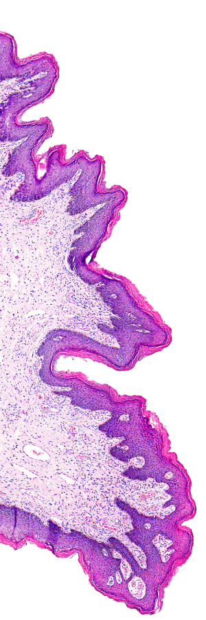
Imagine, if you will, a left-handed bicycle tire, a right-handed fork or a tumor board without, well, pathology images of the tumor.
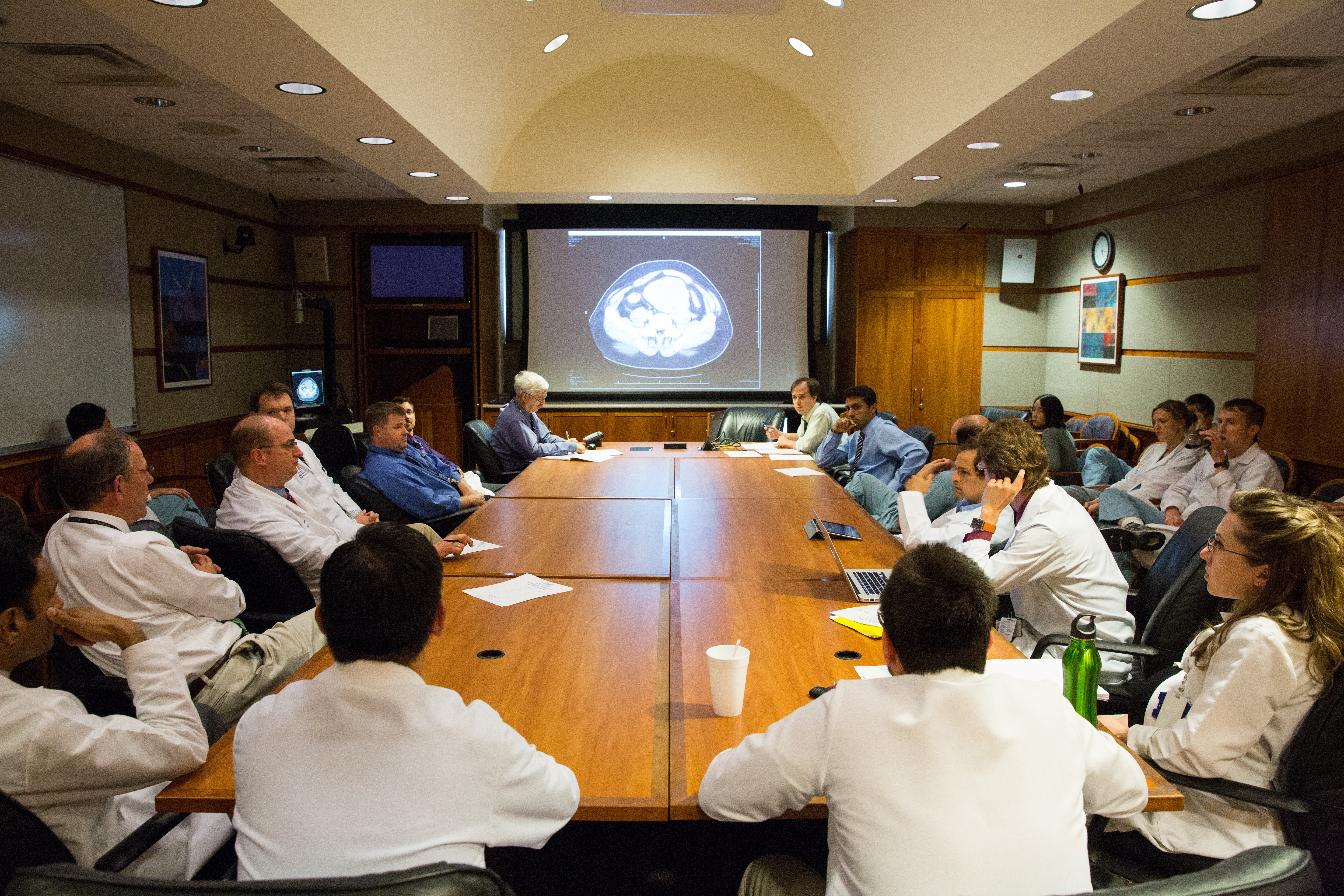
Tumor boards are a multi-disciplinary conference where patients (cases) are presented – their clinical history, radiology findings, and pathology findings (whether biopsy or resection material) – are presented to clinicians for discussions on treatment, management, and best patient care.
Radiologists use PACS systems. Pathologists historically have used video cameras on microscopes, PowerPoint slides of static images to show relevant findings, or whole slide images where clinicians would rather not have conference than do so with anything short of whole slide images.
News from Springfield Memorial from a young pathologist who commented that her tumor boards are without tumor images…pathology images that is. Radiology images are shown but the leader of the tumor board gave up on looking at pathology slides “a long time ago”. So, she shows up to the tumor board, not with slides in hand like so many before her, or with a thumb drive with a slide presentation, or a login for a whole slide image viewer on the hospital network, but with a stack of pathology reports to read to the clinicians. The reports are also available and can be read from the PACS system as well as the EHR and often times are read off the PACS by the radiologist in terms of the pathology findings.
Does anyone else have this in his or her practice?
First and foremost, tumor boards are an integral part of an established Quality Assurance/Quality Control/Performance Improvement (QA/QC/PI) program. Re-reviewing of cases prior to clinical discussion allows another opportunity to present any additional findings not previously mentioned, or perhaps in some cases modify the diagnosis/stage/etc., prior to treatment being initiated, after tumor board discussion.
Secondly, as much as the radiology images are reviewed with the group, pathology images are usually also shared in terms of work-up and steps in the process used to make the diagnosis. Were additional sections taken? Recuts? Immunohistochemistry? Internal review? Outside consultation?
This also allows for a longitudinal review of the patient’s pathology – the original fine needle aspiration, subsequent core needle biopsy, lumpectomy and sentinel lymph biopsy, for example.
Reading the most recent report accomplishes none of this.
Unfortunately, this is an all but too uncommon scenario for many pathology groups/laboratories.
The first and last time I personally experienced a tumor board without pathology image review was more than 10 years ago while serving as the consulting pathologist/medical director for a small laboratory many miles away without a full-time pathologist.
At the time “tumor boards” consisted of the consulting pathologist reading pathology reports over speaker phone to the remote laboratory.
The surgeon who headed up the tumor board and myself, when we took over this activity, changed all of this by implementing video teleconferencing and screen sharing with either live video microscopy and/or static images of gross/histology images.
Other hospitals in the network wanted to replicate or join in on the video virtual tumor board experience.
Folks became more engaged, including pathologists who offered to present at the tumor boards, share their cases and discuss cases with the image as the substrate for discussion.
Curious to hear from others. How many conduct tumor boards at your respective organizations/institutions without showing representative images or slides?
Should this be a requirement for a tumor board? Or are slides/images simply not necessary?
Dr. Kaplan blogs daily at the Digital Pathology Blog/TissuePathology.com and expects the Chicago Blackhawks to win the Stanley Cup, again, this year.
