Digital Pathology Blog

Blog.jpg

Posted by Robin Weisburger 08/08/2023

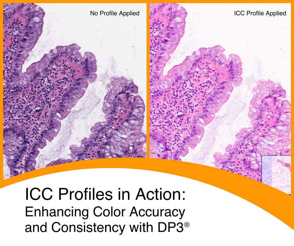
International Color Consortium: Enhancing Color Accuracy and Consistency for Your Digital Pathology Images

The ICC Profile: What Is It and Why Is It Important When Considering Digital Pathology Technologies?

Consistency and reliability of image quality is more important than ever as the adoption of digital pathology increases across clinical, education and research workflows.

The International Color Consortium (ICC) has developed a set of standards to be applied in the management of color for digital images. In color management, the ICC profile is a set of data that characterizes a color input or output device or a color space according to standards promulgated by the Consortium.

Topics: Digital Pathology, Pathology, Slide Management

Posted by David C. Wilbur, M.D. 04/05/2022

Proficiency Testing in the Digital Pathology Age

Don’t get me wrong based on the title; I am not advocating for proficiency testing (PT). After Medical Boards, Resident In-Service and Board exams, I never thought I would be thinking about any more testing at all. There are better ways to test for ongoing real proficiency. Exercises that test “real-world” skills, such as focused review of signed out cases or concurrent reviews - with feedback – not only lead to better outcomes (patient safety) but also improve practice (constructive feedback). But testing is an unfortunate part of modern life. It’s a metric, and regulators like metrics. For years we dealt with the specter of impending gynecologic cytology PT. Based on the complexity of the federally mandated glass slide testing format, the organizations capable of producing such a monumental effort appeared to reach a détente with CMS, and no testing took place for many years after the federal regulation went into effect. However, when one organization came up with a program, the seeming détente was at an end, and CAP and ASCP were essentially forced to enter the fray.

Topics: Digital Pathology, Pathology, digital imaging, Slide Management

Posted by Corista 10/23/2019

Pathologists: Why Go Scanner Agnostic for Your Image Management System?

The key to unlocking the broad benefits of digital pathology, including more efficient use of resources, easy access to colleagues & experts, faster clinical workflows — depends on having an image management system providing broad access to every image in the repository for users to view and share.

Topics: Slide Management

Posted by Robin Weisburger 03/19/2019

Change Evolution in the Laboratory: IHC to DP

The acceptance of digital pathology and its implementation across the United States have been met with a fair amount of challenges. Many equate digital pathology’s evolution in the laboratory with radiology’s move from film to digital media. Despite obvious advantages like immediate availability of images for viewing and image storage being reduced to computer drives as opposed to rooms full of film, it took time for radiology to convert to using digital media as a routine matter of business. Pathology is experiencing much of the same resistance.

Topics: Slide Management

Posted by Robin Weisburger 12/11/2018

Part 4: Challenges to providing a digital pathology service at an international level

This article is the fourth in a four-part series highlighting the evolution of digital pathology and its impact on the access to pathology services throughout the world. If you missed it you can find Part 1 here, Part 2 here, and Part 3 here.

Topics: Slide Management

Posted by Robin Weisburger 12/04/2018

Part 3: A Timeline of Global Pathology Initiatives

This article is the third in a four-part series highlighting the evolution of digital pathology and its impact on the access to pathology services throughout the world. If you missed it you can find Part 1 here and Part 2 here.

Topics: Slide Management

Posted by Robin Weisburger 11/13/2018

Part 2: The Rise and Role of Telepathology

This article is the second in a four part series highlighting the evolution of digital pathology and its impact on the access to pathology services throughout the world. If you missed it, you can find Part 1 here.

Many pathology practices associated with academic medical centers have begun using digital pathology to facilitate the sharing of cases through the use of internet services, private networks, and now, cloud technology. As the use of whole slide imaging (WSI) and digital pathology consultations becomes more widespread, hospitals can access experts in any pathology subspecialty from any location around the globe. Secure, web-based access to expert centers and remote subspecialist colleagues is now a reality.1

Topics: Slide Management

Posted by Robin Weisburger 11/06/2018

Part 1: Doing More With Less — Changing the Face of Pathology

This article is the first in a four part series highlighting the evolution of digital pathology and its impact on the access to pathology services throughout the world.

"In 2012, there were 14 million new cases of cancer reported and 8.2 million cancer-related deaths worldwide. The number of new cancer cases will rise to 22 million within the next two decades.”1

With cancer among the leading causes of death worldwide, the demand for pathology services is becoming more critical. However, the number of new pathologists entering the field is decreasing while the projected demands of the patient population are expected to grow by more than 150% over the next 20 years.

Topics: Slide Management

Posted by Keith Kaplan, MD, Chief Medical Officer 10/16/2018

The Evolution of Reporting: Are Pathologists Becoming Data Managers?

25+ years ago, as a resident, the hard(er) part was looking at the slides and the easy part was writing the report. Before breast conserving therapies, when mastectomies were standard of care, for example, a report might read: “Breast, right, mastectomy: Invasive mammary carcinoma, grade 3, measuring 3.4 cm; deep margin and nipple negative for tumor.” With the header, all the necessary elements in the report, the logo, gross description and disclaimers you could get this onto 1 page. And have space left over. With large fonts (the initial MUMPS based laboratory information systems did not give you the option of 500+ font styles and types with bold, italics, etc…)

Topics: Slide Management

Posted by Corista Marketing 08/09/2016

Digital Study Sets Come of Age

During a recent family dinner our 17-year old was discussing her post-high school plans, which led to a walk down memory lane for her parents. While sharing our own ”ancient” college experiences, she was intrigued (rather than horrified) to learn that I had spent the lion’s share of my last year as an undergrad in the morgue gathering specimens at autopsy. Seems old moms can be cool after all!

During my senior year, we were running a study (using specimens from posts) to search for a protocol that might lead to a pre-symptomatic diagnosis of pancreatic carcinoma – particularly for tumors located away from the duct, in the body or tail. Even then, finding a way to diagnose pancreatic CA “early” was the Holy Grail – and strongly correlated with a vastly better survival rate.

My senior thesis required the collection of dozens and dozens of pancreatic FNAs, which meant hundreds of smears were created and stained. In those long-ago days, this translated into box loads of unwieldy, fragile glass slides needing a permanent place where they could be organized and filed for future retrieval – slides that must be kept carefully segregated from those generated in our clinical lab. In addition, demographic data needed to be “attached” to correlate with microscopic findings. In short, a “study set” was created for each autopsy. In the dark ages of pathology, study sets consisted of a clear plastic “holder” containing a large sleeve to hold patient data on a 3” by 5” card, and four slender sleeves to hold glass slides.

Over time, there was inevitable attrition among our study sets. The plastic holders would crack as they aged. Irreplaceable slides would become broken. If not stained or coverslipped properly, slides would dry out or fade over time. Other slides might be borrowed for publication, presentation, teaching, or further study, and never returned to their sleeve. Often we would pull a much-needed study set, only to find a plastic sleeve containing a sole index card with no slides.

Fast-forward to today, and the valuable “study set” is now a dynamic, searchable, easily accessed, portable, sharable, and reproducible resource – no longer limited by the boundaries of geography or sample size or type of preparation. The advent of digital slides has transformed this cornerstone of pathology, making the creation of unlimited virtual study sets both simple and affordable. When incorporating searchable, text-based functionality across case metadata (tissue description, grossing data, demographics, patient history, and reports), pathologists gain a powerful tool for creating, annotating, sharing, storing, and coordinating cases for publication, training, and clinical utility. A few areas where digital study sets are of particular use include:

Correlation
Digital study sets seem tailor-made to document correlation in a wide array of comparative situations; histo-cyto, colposcopic-histo, and gross-micro. With the press of a button, images are quickly available for comparison – whether for QA, research, publication, teaching, or to build a more complete, robust diagnostic picture.

Changes over time
Watching a patient over time is critical when evaluating certain conditions. A good example is when patients undergo solid organ transplant. Over time, repeat serial biopsies are taken to assess for graft rejection and the health of the graft, to see if the primary disease has recurred. Digital slides make it possible to review all the former prior biopsies quickly and efficiently, and in context of the most recent biopsy.

Prognostic picture
Building prognostic study sets goes beyond the basic H&E or Pap stain. The ability to easily include immuno and molecular stains, photos of gross specimens, and even radiologic images, provide a more comprehensive picture upon which a tailored therapeutic protocol can be built. For a breast cancer patient, this might include several H&Es of the tumor (if the grading is heterogeneous throughout), along with immuno stains for ER, PR, and HER2 – and even a cytokeratin cocktail stain for micromets in an axillary node – all visible on a single screen.

Temporary preparations
Immuno and molecular fluorescent stains can vanish in the blink of an eye. Whether for clinical documentation, publication, or training, building digital study sets of these ephemeral slides provide the shelf life missing in their glass counterparts.

Training and test sets
When learning to diagnose rare lesions – or common ones for which there are challenging differential diagnosis – the only way to gain proficiency is extensive repetition. The ability to duplicate and share digital study sets removes any barrier to access, easily encouraging repeated exposure.

Recently, I uncovered a dusty archive box with the intent of going through the contents in preparation for disposal. It had been buried in the back of my office storage closet for decades, so I was fairly certain nothing precious was inside, but what a sight awaited me. It was filled to the brim with 35mm slides still loaded in their carousels, and old glass-based study sets stored in plastic sleeves. Not quite buggy whips, but antiques nonetheless. Using today’s digital slides, the entire box would fit into one small corner of a reproducible, pocket-size, multi terabyte drive, with room left over for plenty more!

Topics: Slide Management